
1. 정확한 진단을 받고 반려견의 요로결석 유형에 따른 영양 지침을 따르세요.
2. 개가 요산염 결석을 일으키기 쉬우면 집에서 만든 저퓨린 식단으로 전환하는 것이 좋습니다.
3. 결석 형성을 예방하는 데 불필요하거나 효과가 없는 저단백 처방 식품을 피하십시오.
4. 계속해서 결석이 생기는 수컷 강아지의 경우 요도 삽입술 수술을 고려하여 폐쇄 위험을 크게 줄이십시오.
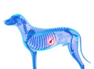
송곳니 신장 결석과 방광 결석은 고통스럽고 생명을 위협할 수 있지만 정보에 입각한 간병인이 이를 예방하는 데 도움이 될 수 있습니다. 지금까지 개에서 가장 흔한 요로 결석 또는 결석은 스트루바이트("개 신장 결석 및 방광 결석 예방" 참조) 및 칼슘 옥살레이트 결석("개에서 방광 및 신장 결석 예방" 참조)입니다. 이 두 가지 유형은 전체 송곳니 요로결석의 약 80%를 차지합니다.
이제 우리는 가장 친한 친구에게 영향을 미칠 수 있는 나머지 결석인 요산염, 시스틴, 인산칼슘, 실리카, 크산틴 및 혼합 또는 복합 요석에 대해 설명합니다.
나머지 돌 범주 중에서 요산염 또는 퓨린 결석이 가장 일반적입니다. 요산암모늄, 요산나트륨 또는 요산을 함유하고 있습니다.
모든 요로 결석의 6~8%만이 요산염 또는 퓨린 결석이지만 특정 품종에서는 그 존재가 상당합니다. 달마시안, 잉글리시 불독, 러시안 블랙 테리어, 라지 먼스터랜더는 유전적 대사 이상으로 인해 요산염이 발생합니다. 미니어처 슈나우저와 요크셔 테리어는 간을 우회하는 비정상적인 혈관인 문맥전신 단락(portosystemic shunt)을 갖는 경향이 있기 때문에 그렇게 합니다. 이 결석은 아주 어린 강아지부터 노인에 이르기까지 모든 연령의 개에서 형성될 수 있지만 요산염이 형성되는 가장 일반적인 연령은 1~4세입니다.

요산염 결석이 생기는 품종 중에서 달마시안이 가장 큰 영향을 받습니다. 1981년에서 2000년 사이에 미네소타 대학 수의과 대학의 미네소타 요로 결석 센터는 달마시안의 돌 7,560개를 분석했습니다. 이 중 97%는 남성이고 95%는 요산염으로 구성되어 있습니다. 수컷 달마시안의 27~34%가 요산염 결석을 형성하는 반면 암컷의 발병률은 훨씬 낮은 것으로 추산됩니다.
달마시안이 형성하는 모든 돌이 요산염이라고 가정하는 것은 유혹적이지만, 수컷 달마시안의 돌 중 97%가 요산염이었지만 소량의 스트루바이트, 크산틴, 옥살산칼슘, 시스틴, 인산칼슘, 실리카 및 혼합 또는 복합도 포함되었습니다. 돌. 암컷 달마시안에 의해 형성된 요석은 69%의 요산염과 29%의 혼합 또는 복합, 2%의 스트루바이트와 0.7%의 잔틴이었습니다. 정확한 식별은 달마시안을 포함한 모든 품종의 요로 결석을 치료하고 예방하는 중요한 첫 번째 단계입니다.
요산염 결석 형성의 주범은 식물 및 동물 조직의 뉴클레오티드 및 핵산에서 발견되는 유기 염기 유형인 퓨린입니다. 식이 퓨린이 분해되면서 요산을 형성하는데, 이는 요산을 형성하는데, 이는 극심한 고통을 주는 관절염 형태인 통풍과 관련이 있는 것으로 인체 의학에서 가장 잘 알려져 있습니다. 민감한 개에서 퓨린은 요산 요로 결석의 형성을 유발합니다.
요산염 결석은 방사선 투과성(즉, 복부 엑스레이로 식별할 수 없음)이므로 종종 초음파, 조영제 엑스레이를 사용하거나 요 결정 또는 채취하거나 제거한 결석을 분석하여 진단합니다.
이전 기사에서 언급했듯이 한 유형의 결석을 예방하거나 치료하는 것이 실제로 다른 유형의 결석을 유발할 수 있으므로 정확한 진단이 필수적입니다. 돌의 정체를 확인하는 유일한 방법은 분석을 받는 것입니다. 그러나 수의사는 소변 pH를 기반으로 교육받은 추측을 할 수 있습니다. 개의 나이, 품종 및 성별; 비뇨기 결정의 확인; 방사선(X-선) 밀도; 감염 여부; 및 특정 혈액 검사 이상.
수의사는 언제 참여해야 합니까? 증상을 발견하자마자 또는 강아지의 품종이 결석이 생기는 경향이 강한 경우에는 더 빨리. 모든 방광과 신장 결석이 위험한 것은 아닙니다. 일부는 여전히 크기가 작지만 배뇨 중에 플러시되고 다른 일부는 신장이나 방광에서 눈에 띄지 않게 남아 있습니다. 결석은 배뇨를 방해할 때까지 합병증을 일으키지 않습니다. 배뇨 곤란, 소변에 피 또는 고름, 고통스럽거나 어려운 배뇨, 배뇨 빈도 증가, 소량의 소변 배출, 평소보다 더 많은 생식기를 핥는 등의 요로 결석 증상에 익숙해지는 것이 중요합니다. ” 집에서 훈련받은 강아지의 경우, 또는 허리의 불편함
긴장한 다음 다량의 소변을 배출하는 개는 방금 돌을 지나갔을 수 있으므로 검사를 받아야 합니다. 돌을 찾을 수 있으면 정확하게 식별할 수 있도록 가지고 가십시오. 소변을 볼 수 없는 개는 요도가 막히면 소변이 시스템으로 역류하여 방광이 파열되거나 신부전이 발생할 수 있으므로 즉각적인 치료가 필요합니다. 늘어난 방광은 근긴장도를 잃어 완전히 비우기가 어려워 감염이나 결석이 생길 수 있습니다. 방광 결석은 암컷의 요도가 더 짧고 넓기 때문에 수컷보다 암컷에서 폐색을 일으킬 가능성이 훨씬 적습니다.
소변량을 늘리고 소변을 볼 수 있는 기회는 모든 유형의 요로 결석을 예방하는 데 중요한 요소입니다. 개가 더 많이 마시고 더 자주 소변을 볼수록 소변 농도가 낮아지고 결석이 될 수 있는 결정이 형성될 가능성이 줄어듭니다. 개가 음식에 물을 추가하고 일반 물 외에 맛이 첨가된 물을 제공하여 더 많이 마시도록 격려하십시오. 요산염 결석이 있는 강아지의 경우 소금을 추가할 수 있습니다.
갈증을 증가시키기 위해 음식에 첨가합니다(꼬집음부터 시작하여 개의 반응을 관찰하고 개가 더 많은 물을 마실 때까지 조금씩 추가). 그러나 시스틴, 인산칼슘 또는 규소 결석을 형성하기 쉬운 개에게 소금을 추가하는 것은 피해야 합니다.
개가 소변을 오랫동안 참아야 하는 경우 소변이 과포화 상태가 될 가능성이 더 높아져 미네랄이 결정으로 침전되기 시작하기 때문에 개가 소변을 볼 기회가 자주 있는지 확인하십시오.
요산염 형성 개를 건강하게 유지하는 열쇠는 저퓨린 식단을 먹이는 것입니다. 요산 결석 형성을 유발하는 퓨린이 없으면 민감한 개라도 정상적인 삶을 영위할 수 있습니다.
일부 달마시안 주인은 결석이 생기기 쉬운 개에게 미네랄이 없는 증류수만 주는 것이 결석이 더 많이 생기는 것을 방지하는 데 도움이 된다고 믿습니다. 그러나 이에 대한 과학적 증거는 없습니다. 강아지가 섭취하는 물의 양이 미네랄 함량보다 더 중요할 수 있습니다.
요산염 결석은 산성 소변에서 발생하기 때문에 추가 예방 전략은 알칼리화 효과가 있는 음식을 먹이는 것입니다. 일반적으로 육류는 산성화 식품이지만 대부분의 과일과 채소는 알칼리화 효과가 있습니다. 이러한 이유로 채식 개 사료가 권장되기도 하지만 채식은 불완전하다고 생각합니다. 또한 단백질 공급원으로 콩을 사용하는 음식은 콩에 퓨린이 많기 때문에 요산염 결석이 생기기 쉬운 강아지에게 적합하지 않습니다. 그러나 콩이 없는 채식 식품은 계란, 요구르트, 치즈 및 기타 저퓨린 단백질 공급원이 추가되는 기초로 사용될 수 있습니다.
Sojo의 Grain Free Dog Food Mix와 같은 일부 개 사료 사전 혼합도 마찬가지입니다. Sojo's Complete는 고구마, 칠면조 및 계란을 기본으로 하며 고요산혈증(소변에 과도한 양의 요산)이 있는 개에게도 적합할 수 있습니다. 알팔파, 귀리, 보리 또는 기타 퓨린 함량이 높은 식품이 많이 포함된 혼합물을 피하십시오("다양한 식품의 퓨린 함량" 참조).
요산염 결석은 저퓨린 식이, 소변 알칼리화 및 2차 감염 조절의 조합으로 용해될 수 있습니다. 용해 중 소변 pH의 목표 범위는 7.0~7.5입니다. 너무 많이 알칼리화하여 소변 pH를 7.5보다 높게 만들지 않도록 주의해야 합니다. 이는 인산칼슘 결석이나 요산염 결석 주위에 껍질을 형성하여 용해하기 어렵거나 불가능하게 만들 수 있기 때문입니다.
크산틴 산화효소 억제제 알로퓨리놀(상품명 Zyloprim)은 결석을 용해하는 데 도움이 될 수 있는 개의 요산 생성을 줄이거나 억제하기 위해 단기적으로 처방될 수 있습니다. 이 약은 문맥전신 단락이 있는 환자에게 사용해서는 안됩니다. 알로퓨리놀을 투여하는 동안 낮은 퓨린 식단을 제공해야 합니다. 그렇지 않으면 개가 크산틴 결석과 껍질을 형성하여 용해를 어렵게 만들기 때문입니다. 예방으로 알로퓨리놀을 장기간 사용하는 것은 권장되지 않지만 다른 치료에도 불구하고 문제가 지속되는 경우 저용량으로 고려할 수 있습니다.
평균적으로 알로퓨리놀을 저퓨린 식이 및 요 알칼리화와 함께 사용하면 결석이 용해되는 데 약 3개월 반이 걸리지만 짧게는 한 달에서 길게는 18개월까지 걸릴 수 있습니다. 결석이 작아지면 요도로 이동하여 폐색을 유발할 수 있습니다.
요산암모늄으로 추정되는 심각한 신장 결석의 일부 사례는 수술 션트 교정만으로 저절로 해결되었습니다.
요산 결석이 생기기 쉬운 개의 pH를 중성(7.0)으로 유지하기 위해 테스트 스트립을 사용하여 소변 pH를 모니터링할 수 있습니다. 테스트 스트립은 소변 흐름에 보관할 수 있으며 소변은 테스트를 위해 종이컵, 그릇 또는 기타 용기에 수집할 수 있습니다. 소변을 수집하면 개가 통과할 수 있는 작은 돌이나 거친 "자갈"뿐만 아니라 혈액, 고름 또는 기타 감염 징후를 확인할 수 있습니다. 권장되는 검사 시간은 아침에 가장 먼저 수유 전입니다.
소변 pH의 변화는 결석의 존재 여부를 나타내지 않지만 결석 생성을 유발할 가능성이 다소 있는 상태를 나타내며 식이 변화가 개의 pH에 미치는 영향을 보여줍니다. pH가 갑자기 상승하면 박테리아 감염을 알릴 수 있으므로 의료 조치가 필요합니다. 결석이 생기기 쉬운 개의 요로 감염을 조절하는 것이 중요합니다. 소변이 산성으로 남아 있고 결정뇨(요 결정 형성)가 지속되면 구연산칼륨 또는 중탄산나트륨과 같은 알칼리화제를 추가할 수 있습니다.
고요산혈증은 높은 수준의 요산이 배설되어 요산염 결석이 형성되는 것이 특징입니다. 고요산뇨증을 유발하는 결함 유전자가 데이비스 캘리포니아 대학의 연구원들에 의해 발견된 후, 질병과 관련된 돌연변이를 탐지하기 위한 테스트가 개발되었습니다. 이 테스트는 모든 품종에 유효합니다.
고요산혈증의 영향을 받은 개는 각 부모로부터 하나씩 유전되는 두 개의 돌연변이 사본을 가지고 있습니다. 돌연변이 사본이 한 개만 있는 개는 자손의 평균 50%에게 돌연변이를 전달하는 증상이 없는 보인자입니다. 육종가는 DNA 테스트를 사용하여 보인자를 식별하고 달마시안 이외의 품종에서 고요산뇨증을 효과적으로 근절할 수 있습니다. (현재 미국에 등록된 모든 달마시안이 돌연변이의 영향을 받습니다. "LUA 달마시안"을 참조하세요. 어미와 부계 모두 돌연변이가 제거되면 그들의 강아지도 모두 제거됩니다.
DNA 검사는 3가지 범주로 개를 식별합니다. 고요산뇨증과 함께(개에는 2개의 돌연변이 사본이 있어 요산 결석 장애로 이어질 수 있는 높은 산 수치를 유발함).
고요산혈증에 걸린 모든 개는 잠재적인 요산염 결석 형성자입니다. 언제든지 고 퓨린 식품, 불충분한 수분, 불충분한 배뇨 기회, 지나치게 산성인 소변의 조합으로 인해 요산 요로 결석이 형성될 수 있습니다. 요산 결정을 확인하기 위한 정기적인 일상 소변 검사는 고요산뇨증이 있는 개를 모니터링하는 데 사용할 수 있습니다. 이 목적을 위한 가장 정확한 샘플은 개가 밤새 소변을 보지 않았다고 가정하고 아침에 수집하므로 소변이 더 농축됩니다. 샘플은 깨끗한 유리, 플라스틱 또는 기타 화학적으로 불활성인 용기에 수집해야 합니다. 잘못된 결정화를 방지하려면 샘플을 냉장 보관하지 말고 30분 이내에 또는 가능한 한 빨리 테스트해야 합니다.
많은 달마시안은 결코 돌을 생성하지 않지만, 돌을 생성할 수 없다고 가정하는 것은 안전하지 않습니다. 널리 보고된 한 사례에서 증상을 전혀 나타내지 않은 13세 달마시안이 하루에 두 숟가락의 새로운 보충제를 받기 시작했습니다. 그 이전에 그의 식단은 그의 성인 생활 내내 동일했습니다. 몇 주 안에 그의 요로가 요산염 결석에 의해 완전히 막혔습니다. 보충제에는 단백질이 적었지만(14%만) 단백질 공급원은 고퓨린 식품인 간이었습니다.
음식에서 퓨린을 줄이는 것은 요산염 결석의 위험을 줄이는 효과적인 방법입니다. 대부분의 고단백 식품은 퓨린 함량이 높기 때문에 수의사들은 종종 요산염 형성 개를 저단백 식단으로 전환할 것을 권장합니다. 그러나 요산염 문제를 일으키는 것은 단백질의 양이 아닙니다. 일종의 단백질이다. 달마시안과 다른 요산염 경향이 있는 개는 퓨린이 적은 단백질이 풍부한 식단에서 번성하는 반면, 이 같은 개는 소량의 고 퓨린 성분이 포함된 저단백 식품을 섭취한 후에도 결석이 생길 수 있습니다. 저단백 식단은 성견에게 장기간 먹일 경우 영양 결핍을 유발할 수 있으며, 강아지와 임산부 또는 수유 중인 여성에게는 전혀 적합하지 않습니다. (아래 "저단백 다이어트의 부작용"을 참조하십시오.)
영양 결핍 없이 퓨린 함량이 낮은 상업용 애완 동물 사료를 찾기가 어렵기 때문에 많은 요산염 형성 개 소유자가 집에서 만든 식단을 먹입니다. 전 세계의 개 애호가들에게 BARF(뼈와 생식 또는 생물학적으로 적합한 생식) 다이어트를 소개한 책 Give Your Dog a Bone을 쓴 호주 수의사 Ian Billinghurst는 여러 웹사이트.
그는 이렇게 말합니다. “오늘날 서구 국가에서는 석재 형성자를 위한 일반적인 가정식 식단에 약 80%의 쌀, 10%의 야채, 5%의 고기가 포함될 것이라고 믿게 되었습니다. 이것은 어떤 개에게나 먹일 수 있는 끔찍한 식단입니다. 이것은 그것을 견디도록 강요된 개들에 의해 입증됩니다. 그들은 계속되는 굶주림, 에너지 부족, 열악한 코트 상태, 체중 유지의 어려움 또는 심각한 체중 감소를 포함하여 수많은 문제로 고통 받고 있습니다.” 이러한 식단은 단백질, 지방, 비타민, 미네랄이 부족할 뿐만 아니라 결석 형성을 예방하지 못합니다.
Billinghurst 박사가 추천하는 생고기 뼈는 닭 목, 닭 등뼈, 닭 날개, 칠면조 목입니다. "잎이 많은 채소를 많이 포함하여 퓌레 또는 펄프 야채를 많이 사용하십시오."라고 그는 말합니다. 식단에는 계란, 코티지 또는 리코타 치즈, 요구르트, 올리브 또는 아마씨 기름이 포함될 수 있으며 비타민 B 복합체, 비타민 E, 다시마, 대구 간유 1티스푼이 일주일에 여러 번 보충됩니다.” 대구 간유는 간을 포함하지 않는 집에서 만든 식단을 먹인 요산염 생성 개에게 중요합니다.
변화하는 다양한 계란, 치즈, 유제품, 소량의 중간 퓨린 육류, 가금류 및 생선과 퓨린 함량이 낮은 채소, 과일 및 보조제와 소변을 희석할 수 있는 충분한 물을 급여하면 요산염을 형성하는 개는 건강하고 행복합니다.
시스틴은 피부, 모발, 뼈 및 결합 조직의 건강에 필수적인 황 함유 아미노산입니다. 과잉 시스틴은 일반적으로 신장에서 걸러져 소변으로 들어가지 않지만 일부 개는 이러한 걸러내는 작용을 방해하는 유전 대사 장애인 시스틴뇨증을 가지고 태어납니다. 시스틴이 소변으로 들어가면 결정체와 요로결석을 형성할 수 있습니다.
시스틴 결석은 드물며 실험실에서 확인된 요로 결석의 1% 이하를 나타냅니다. 모든 품종에서 시스틴뇨증이 발생할 수 있지만 특정 품종이 가장 영향을 받습니다. 남성 마스티프의 약 10%가 시스틴뇨증을 가지고 있습니다. 뉴펀들랜드, 잉글리시 불독, 스코티시 디어하운드, 닥스훈트, 스태퍼드셔 불 테리어, 치와와에서도 흔히 볼 수 있습니다. 시스틴 결석은 방사선 불투과성이 약하여 칼슘이 포함된 결석보다 X-레이에서 보기가 더 어렵습니다.
시스틴뇨증에는 적어도 두 가지 유형이 있습니다. 더 심각한 형태는 뉴펀들랜드와 드물게 래브라도 리트리버, 그리고 아마도 다른 일부 품종과 믹스에 영향을 미칩니다. 이 개에서는 수컷과 암컷이 동등하게 영향을 받습니다(항상 그렇듯이 수컷이 폐쇄될 가능성이 더 높음). 발병 연령은 6개월에서 1세까지 젊을 수 있습니다. 수술 후 결석의 재발은 이러한 개에서 더 빠르며 신장 결석을 형성할 가능성이 더 높습니다. The gene that causes cystinuria in these breeds has been identified and a simple, reliable genetic test can identify both affected dogs and carriers.
In other breeds, dogs with cystinuria are almost always male. No genetic test is available for them, though the University of Pennsylvania School of Veterinary Medicine (PennVet) is collecting blood samples from affected Mastiffs and their genetic relatives to try to produce a DNA test. The average age at onset of clinical signs is about 5 years.
A basic urinalysis can sometimes detect cystine in urine, though this is the least reliable method of detection. A nitroprusside (NP) test performed at the University of Pennsylvania (PennGen) is considered more reliable. A quantitative amino acid analysis performed by PennGen or a human medical laboratory is most reliable but very expensive. If cystine is found in the urine on any of these tests, the diagnosis is considered positive for cystinuria, though that doesn’t necessarily mean the dog will form stones.
Unfortunately, a negative result on any of these tests does not guarantee that the dog is “clear.” Note that sulfa drugs and supplements, including sulfa antibiotics, MSM, and Deramaxx, may cause false positive results.
“Cystinuria is a particularly frustrating condition to manage,” says San Francisco Chronicle pet columnist Christie Keith, who started a Canine Cystinuria e-mail list and website when one of her Scottish Deerhounds developed cystine uroliths. “A dog known to have cystinuria may go his whole life without obstructing, while another dog, never diagnosed, can have a life-threatening obstruction as his first symptom. It’s not known at this time why some dogs with cystinuria form stones and others do not.”
Cystine, like all amino acids, is one of the building blocks of protein. That’s why most veterinarians (including many kidney specialists) prescribe a low-protein diet, speculating that reducing the cystine supply will reduce the formation of cystine stones. Another common recommendation is to alkalize the dog’s urine because cystine stones form in acid urine.
Unfortunately, says Keith, these strategies are ineffective. “Most of us on the Canine Cystinuria list have found that diet and urinary alkalization have failed to prevent our dogs from forming stones,” she says, “and they have sometimes caused other problems, including other types of stones that form in alkaline urine. If the urine goes into acidity even briefly, cystine stones can form and they won’t dissolve just because alkaline urine is achieved soon after. In addition, feeding ultra-low-protein diets can be dangerous, especially to giant breeds and breeds prone to cardiomyopathy.” (See “The Side Effects of Low-Protein Diets”, below.)
It’s important to provide your dog with extra fluids and frequent opportunities to urinate in order to keep his urine from becoming supersaturated. Salt should not be added to increase fluid consumption for dogs with cystinuria; according to studies conducted on humans, a low-sodium diet may decrease the amount of cystine in the urine.
If urine alkalization is attempted, the target pH is 7.0 to 7.5; higher can predispose dogs to calcium phosphate uroliths. Potassium citrate is preferred for alkalization when needed rather than sodium bicarbonate because sodium may enhance cystinuria.
Cystine stones cannot be dissolved with diet or supplements, but two prescription drugs can help dissolve and prevent them. Cuprimine (d-penicillamine) has potentially serious side effects but is less expensive and more readily available, and many dogs do well on it. According to Keith, Thiola (tiopronin, also referred to as 2-mercaptopropionylglycine or 2‑MPG), has fewer side effects, but one of them is the depletion of the owner’s bank account. Maintaining a giant-breed dog on Thiola can cost as much as $500 per month. Because the severity of cystinuria tends to decline with age, the dosage of preventative medications can sometimes be decreased or even stopped.
Dissolution requires a combination of medication, low-protein diet, and urinary alkalinization. Even then it may not be successful or practical for a dog with numerous stones. When it does work, dissolution commonly takes one to three months.
For some dogs, the solution has come not from prevention strategies or medication but from surgery. “It sounds extreme,” says Keith, “but many of us who have stone-forming male dogs with cystinuria have opted for a scrotal urethrostomy. This surgery redirects the dog’s urethra away from the penis to a new, surgically created opening in front of the scrotum.”

The wider opening that results enables males to more easily pass small stones and help prevent urinary blockages. “While future obstruction is not impossible,” says Keith, “this procedure reduces the risk substantially.” Still, she cautions, this surgery should not be undertaken lightly. It’s expensive, requiring the expertise of a skilled board-certified surgeon, and because the affected area is rich in blood vessels, there can be significant post-surgical bleeding, though the surgery is not particularly painful.
“The good news,” she says, “is that many dogs, including stone-formers and those who had serious complications when their condition was first diagnosed, have lived not just normal but longer-than-normal lives.”
Like cystine stones, stones composed of xanthine, calcium phosphate, and silica are rare, each representing less than 1 percent of analyzed uroliths. Ironically, they often occur while the patient is undergoing treatment for the prevention of other stones.
– Although xanthine is a type of purine, xanthine stones are associated not with diet but with the use of allopurinol. Xanthine crystals almost never occur naturally, though they have been reported in some cats, Cavalier King Charles Spaniels, and Dachshunds. The average age at onset is 6 to 7 years. Like urate stones, they are radiolucent; that is, they cannot be seen on X-rays.
In some cases, discontinuing allopurinol while feeding a low-purine diet has dissolved xanthine uroliths, but in general, treatment consists of surgical removal, urohydropropulsion (a nonsurgical procedure performed with the dog under anesthetic, in which the bladder is filled with saline through a catheter, and the bladder is manually squeezed to force stones out through the urethra), or lithotripsy (the use of high-energy sound waves to break up the stones).
A low-protein diet is usually recommended for dogs receiving allopurinol treatment (to help prevent formation of xanthine uroliths); but again, what’s really needed is a low-purine diet.
– Calcium phosphate stones often develop when the urine is over-alkalized (at a pH greater than 7.5), in an effort to prevent the formation of calcium oxalate, urate, or cystine stones. The average age at onset is 7 to 8 years, but these stones have been found in dogs of all ages, including puppies and seniors.
Calcium phosphate stones are commonly called apatite uroliths, with hydroxyapatite and carbonate apatite the most common. They are radiographically dense, so they are easily seen on X-rays. Uroliths composed primarily of calcium phosphate are rare and associated with metabolic disorders such as hyperadrenocorticism (Cushing’s disease), hypercalcemia, renal tubular acidosis, or excessive calcium and phosphorus in the diet.
Because they cannot be dissolved medically, these stones are usually removed surgically, though that may be unnecessary if the stones are clinically inactive (not growing or causing problems). They have been known to dissolve spontaneously following parathyroidectomy surgery for primary hyperparathyroidism. Unless the patient has a metabolic condition that contributes to calcium phosphate stones, the strategies used for prevention are similar to those used for calcium oxalate stones, although it’s important to avoid excessive alkalization of the urine.
Medications that can enhance calcium excretion, including prednisone and furosemide (Lasix), should be avoided if possible. Salt should not be added to the diet, as sodium increases urinary calcium.
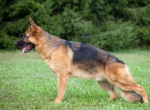
– Silica stones are most common in male German Shepherds, Old English Sheepdogs, Golden Retrievers, and Labrador Retrievers, although other breeds and mixed breed dogs have developed them as well. More than 95 percent of silica stones occur in males. The problem can develop in dogs as young as four months or as old as 12 years, but most stones occur in dogs aged 6 to 9 years. Silica stones are radiopaque and can be seen on X-rays. No relationship has been found between urinary pH and silicate urolith formation.
The formation of silica stones is associated with diets high in cereal grains, particularly corn gluten and soy bean hulls, both of which are high in silicates. Corn gluten and soy bean hulls (also called soybean mill run) are ingredients in low-quality prescription diets and dog foods.
Other foods that are high in silica, and which should be avoided, include the hulls of wheat, oats, and rice (hulls are found in whole grains); sugar beets; sugar cane pulp; seafood; potatoes and other root vegetables; onions (which shouldn’t be fed to dogs, anyway); bell peppers; asparagus; cabbage; carrots; apples; oranges; cherries; nuts and seeds; grains; soybeans; and the herbs alfalfa, horsetail, comfrey, dandelion, and nettles. Bentonite clay, a mineral supplement, is also high in silicates.
Because no drug or diet dissolves silica stones, they may be removed surgically, flushed out with urohydropropulsion, or shattered with lithotripsy; no treatment may be required for clinically inactive stones. Silica stones do not usually recur, but it makes sense to feed a diet that is high in protein from animal sources and low in plant foods, including fiber and bran. As with all stones, keep the urine diluted by increasing fluids and giving your dog frequent opportunities to urinate. Don’t add salt, which is another source of silica.
Dogs who drink water from sources containing sand may develop silica uroliths, so water that contains silica (a primary mineral in sand) should be avoided. In hard-water areas, distilled water is recommended for dogs who form silica stones. Silica stones have also been associated with pica, an eating disorder that causes dogs to eat dirt, rocks, and other non-food items.
Most bladder stones are caused by a single type of mineral. Sometimes a stone consists of two or more minerals in approximately equal proportions, in which case it is called a mixed urolith. These stones are rare, comprising only 2 percent of analyzed uroliths.
A stone that consists of a core mineral surrounded by a smaller amount of a different mineral is called a compound urolith. These make up 10 to 12 percent of analyzed stones. Compound uroliths can sometimes be identified based on differing radiographic density of their stone layers.
Compound uroliths develop when a stone’s environment changes, such as when a struvite stone is treated by reducing urinary pH, magnesium, and phosphorus, resulting in a calcium oxalate shell around the struvite core. Struvite shells caused by infection commonly form over calcium oxalate and other cores, especially since all stones predispose dogs to bladder infections.
One treatment strategy is to try to dissolve the outer layer first. This is especially effective for stones with an infection-induced struvite shell, which make up more than 80 percent of compound uroliths with cores other than struvite. The struvite shell should dissolve with appropriate antibiotic or infection-fighting treatment. X-rays can be used to monitor dissolution. Once the outer shell disappears, treatment strategy switches to the inner core, also called the nucleus, or the stones may then be small enough to remove by urohydropropulsion.
More than half of the compound uroliths analyzed in 2002 by the Minnesota Urolith Center contained a calcium oxalate core, and almost all of these were surrounded by a struvite shell caused by infection. Unlike calcium oxalate uroliths, these compound uroliths were found primarily in female dogs; again, this is because the female dogs’ anatomy makes them more susceptible to urinary tract infections, which play a role in causing struvite stones. Treatment and prevention should be focused on controlling infections and reducing the risk of calcium oxalate stones.
Stones with a struvite core made up amost a quarter of compound uroliths, more than half of which were surrounded by a calcium phosphate shell and most of the rest by a calcium oxalate shell. As is common with infection-induced stones, most of these dogs were female.
Urinary acidifiers can contribute to urinary calcium that leads to the formation of calcium-containing stones. Treatment is the same as for struvites:appropriate medication for the infection and possibly a reduced-protein diet short-term to help dissolve the stones quickly. Urinary acidification is not recommended due to the increased risk of calcium oxalate and calcium phosphate formation.
Small percentages (3 to 5 percent each) of compound uroliths were comprised of the following:
- Silica core. Most of these had a calcium oxalate shell and were found in male dogs. Since both silica and calcium oxalate stones are associated with plant-based foods, diets containing substantial plant proteins should be avoided.
– Calcium phosphate core surrounded by struvite or calcium oxalate shells. These are treated the same way as struvite or calcium oxalate stones.
– Urate core , most of which were surrounded by struvite. Treatment is aimed at controlling the infection along with management of the urate core.
– Compound uroliths with a core or shell of xanthine are treated by discontinuing or reducing the dose of allopurinol.
Sulfa drugs may create a shell around struvite uroliths when used at high doses for prolonged periods, or in dogs with acidic or highly concentrated urine. For this reason, sulfa drugs should be avoided when treating lower urinary tract (bladder) infections, particularly for dogs known to have stones or one of these risk factors.
Preventive treatment should focus on whatever minerals comprised the stone’s inner core. As with all types of stones, increasing fluid intake and opportunities to urinate are recommended. Adding salt to the diet is not recommended, however, as it increases urinary calcium and calcium is commonly found in uroliths.
Without sufficient protein in the diet, protein is pulled from muscles to meet the body’s requirements. Nutritionally inadequate, low-protein diets should never be fed to puppies or dogs who are pregnant or nursing, and they can cause health problems if given to adult dogs for prolonged periods.
According to the Merck Veterinary Manual (9th Edition, 2008), “The signs produced by protein deficiency or an improper protein-to-calorie ratio may include any or all of the
following:weight loss, skeletal muscle atrophy, dull unkempt coat, anorexia, reproductive problems, persistent unresponsive parasitism or low-grade microbial infection, impaired protection via vaccination, rapid weight loss after injury or during
disease, and failure to respond properly to treatment of injury or disease.”
Ultra-low-protein diets such as Hill’s Prescription u/d have been linked to dilated cardiomyopathy (DCM) in English Bulldogs, Dalmatians, and other breeds. Dogs with cystinuria, which predisposes dogs to carnitine deficiency even when a normal-protein diet is fed, are particularly at risk. Some Newfoundland dogs are prone to taurine deficiency leading to DCM even when fed regular commercial diets, especially lamb and
rice diets, though many manufacturers now add taurine to their lamb and rice diets to help prevent this side effect.
According to a study of cardiac function in healthy dogs fed protein-restricted diets published in the American Journal of Veterinary Research in 2001, “Dogs fed protein-restricted diets can develop decreased taurine concentrations…The possibility exists that AAFCO [Association Of American Feed Control Officials] recommended minimum requirements are not adequate for dogs consuming protein-restricted diets. Our results also revealed that, similar to cats, dogs can develop DCM secondary to taurine deficiency, and taurine supplementation can result in substantial improvement in cardiac function.”
Low-protein diets are not needed in most cases to prevent the development of kidney or bladder stones. lf you choose to feed a low-protein diet, you should supplement with carnitine and taurine to help prevent the development of DCM. Dogs with cystinuria may benefit from supplementation even if fed a regular diet. Suggested preventative dosages are 25 to 50 mg L-carnitine and 5 mg taurine per pound of body weight two or three times a day. For example, a 50-pound dog should receive 1,250 to 2,500 mg L-carnitine and 250 mg taurine twice or three times a day. Higher dosages are needed to treat DCM.
You can also add eggs and dairy products to a low-protein diet for dogs with hyperuricosuria to increase protein.
Once your dog’s stones are successfully treated, you’ll want to use the strategies described in this article to help keep them from coming back. Stone-forming dogs can be monitored by their veterinarians with X‑rays, ultrasound, and urinalyses.
Infection-induced struvites can recur in as little as a few days to a few weeks, while calcium oxalate and silica stones may take a few months to recur. Cystine and urate stones can recur rapidly. Some dogs continue to form stones despite diet changes and medical therapy. For them the key is monitoring with radiographic imaging (X-rays or ultrasound) at least every 3 to 6 months (more often to start with and for rapidly recurring types) in order to detect stones while they are still small enough to pass through the urethra using urohydropropulsion or catheter-assisted retrieval.
A final solution for males with recurring stone blockages is urethrostomy surgery, which redirects the flow of urine to avoid its normal narrow passage.
CJ Puotinen is the author of The Encyclopedia of Natural Pet Care and other holistic health books. She lives in Montana, and is a frequent contributor to Whole Dog Journal. San Francisco Bay Area resident Mary Straus has spent more than a decade investigating and writing about canine health and nutrition topics for her website, DogAware.com.
개의 췌장염 경고 징후는 처음에는 항상 명확하지 않거나 덜 심각한 질병으로 오인될 수 있지만 상태는 여전히 생명을 위협할 수 있습니다. 따라서 반려견이 지방이 많은 음식에 탐닉하는 경향이 있거나 유전적으로 췌장염에 걸리기 쉬운 경향이 있는 경우, 반려견에서 이 상태의 증상, 원인 및 치료에 대해 더 알고 싶을 수 있습니다. 전문가 팁 :췌장염은 특히 개가 심한 경우에 치료하기에 매우 비싼 상태가 될 수 있습니다. 질병의 중증도에 따라 진단 및 집중 치료에 수천 달러가 필요할 수 있습니다. 애완 동물 보험은 이러한 비용을 충당
고관절 이형성증은 쉽게 고칠 수 없고 치료 비용이 많이 들고 고통스러울 수 있지만 적절한 관리를 통해 통증을 최소화할 수 있는 유전 질환입니다. 이 게시물에서는 반려견을 건강하고 행복하게 유지하기 위해 취할 수 있는 증상, 치료 옵션 및 예방 조치를 포함하여 고관절 이형성증에 대해 알아야 할 모든 것을 배울 것입니다. 목차: 고관절 이형성증이란 무엇입니까? 개의 고관절 이형성증의 증상 고관절 이형성증은 어떻게 진단하나요? 개의 고관절 이형성증 치료 수술 옵션 대체 의학 치료 고관절 이형성증의 치료 비용은 얼마입니까? 고관절
