송곳니 흑색종은 너무 복잡하고 다양하여(그러나 서로 구별되는) 멜라닌 세포 종양 하위 유형 그룹에 대한 포괄적인 용어로, 때로는 완전히 다른 질병처럼 보일 수 있습니다. 모든 유형의 흑색종의 공통점은 정상적인 멜라닌 세포(멜라닌 생성을 담당하는 세포)가 분열하여 통제 불능 상태로 성장할 때 형성된다는 것입니다.
흑색종은 양성 또는 악성 종양으로 분류됩니다. 다행히도 개에서 발생하는 대부분의 흑색종은 양성입니다. 이 형태의 흑색종은 일반적으로 흑색세포종이라고 합니다. 이 종양은 암이 아니며 일반적으로 암이 되지 않으며 정상 세포의 기능을 방해하지도 않습니다. 그들은 특정 크기에 도달하고 다른 조직을 침범하지 않으면 종종 성장을 멈춥니다. 또한 전이되지 않으며 수술로 제거해도 다시 자라지 않는 경향이 있습니다.
대조적으로, 모든 송곳니 흑색종의 5-7%를 차지하는 악성 흑색종은 매우 공격적이며 중요한 장기로 매우 빠르게 전이할 수 있습니다. 미국에서 매년 약 100,000건의 악성 흑색종이 진단됩니다.
이 암성 종양은 착색된 신체 부위에 형성되는 경향이 있으며 종양은 일반적으로 갈색 또는 검은색이지만 생성되는 멜라닌 수준에 따라 분홍색, 황갈색 또는 흰색으로 나타날 수 있습니다. 이들은 성별에 대한 선호도가 없는 중년에서 노령견(평균 연령 9세)에서 가장 흔히 볼 수 있습니다.
신체의 위치는 이 암의 특정한 생물학적 행동을 결정할 것입니다. 개는 암이 퍼질 때까지 무증상인 경우가 많습니다.
원인
송곳니 흑색종의 원인은 알려져 있지 않지만 연구자들은 이것이 환경적 요인과 유전적 요인의 조합으로 인한 것일 수 있다고 생각합니다. 또한 화학 물질, 스트레스, 외상 또는 특정 부위를 과도하게 핥는 것이 요인이 될 수 있다고 의심됩니다. 세포가 무작위로 증식하도록 촉발되면 세포 분열 중 돌연변이 가능성이 증가하고 악성 세포가 형성될 수 있습니다.
자외선 노출은 인간의 흑색종의 주요 원인이지만, 아니다 일반적으로 보호 모피 코트로 인해 송곳니 형태와 관련이 있습니다.
견종 성향
개의 악성 흑색종은 에어데일스, 블러드하운드, 보스턴 테리어, 치와와, 차우차우, 코커 스패니얼, 닥스훈트, 도베르만 핀셔, 잉글리시 스프링거 스패니얼, 골든 리트리버, 고든 세터 등의 품종이 과도하게 대표되는 강력한 유전적 요소를 반영하는 것으로 생각됩니다. , 아이리시 세터, 페키니즈, 푸들, 로트와일러, 미니어처 및 자이언트 슈나우저, 스프링거 스패니얼, 스코티시 테리어, 티베탄 스패니얼.
이 질병은 또한 검은 개의 발가락이나 발톱 바닥에 나타날 가능성이 더 큽니다. 입안에 색소 침착이 심한 작은 품종의 경우 구강 흑색종의 위험이 증가하는 것으로 보고되었습니다.
진단
개의 악성 흑색종의 진단은 일반적으로 종양의 미세침 흡인물로부터의 세포학 및/또는 생검 및 조직병리학을 통해 이루어지지만 진단하기 어려운 것으로도 알려져 있습니다.
흑색종이 착색되면 병리학자는 일반적으로 샘플에서 멜라닌 과립과 특징적인 세포 형태를 볼 수 있습니다. 멜라닌 세포 종양에 색소 침착이 없고 세포 형태가 매우 다양할 때 어려움이 발생합니다.
생검의 조직병리학적 결과는 암종, 육종, 림프종 또는 골형성 종양과 유사할 수 있습니다. 이 시점에서 면역조직화학(IHC) 표지자(Melan-A, PNL-2, 티로신 반응성 단백질 TRP-1 및 TRP-2)에 대한 특수 염색으로 추가 검사가 필요합니다. 이 스크리닝은 멜라닌 세포를 검출하는 데 매우 민감하고 특이적입니다. 사용되는 치료 프로토콜과 예후를 결정하기 때문에 정확한 진단을 받는 것이 중요합니다.
개의 전반적인 건강을 평가하고 질병의 단계를 결정하기 위한 추가 진단 테스트에는 완전한 혈구 수를 포함할 수 있습니다. 혈청 생화학적 프로파일; 소변검사; 전이의 증거를 찾기 위한 흉부 방사선 사진 및 복부 초음파; 세포가 림프계로 퍼졌는지 확인하기 위해 림프절을 흡인합니다.
구강 형태의 흑색종이 있는 개의 경우, 특히 림프절이 비대해진 것으로 확인되면 복부 림프절, 간, 부신 및 기타 부위의 전이를 확인하기 위해 추가 검사가 필요합니다.
구강 종양의 경우 방사선 사진 및/또는 컴퓨터 단층촬영(CT) 스캔이 권장될 수 있습니다.
디지털(발가락) 흑색종은 종종 뼈 파괴를 수반하기 때문에 영향을 받은 발의 방사선 사진을 찍어야 합니다.
안구 흑색종의 특정 진단 기술에는 세극등 검사, 안압 측정법(안압), 측각경 검사(눈 앞부분 검사) 및 안저검사(눈 뒤쪽 검사)가 포함됩니다.
스테이지
위에서 논의한 진단 테스트는 환자의 악성 흑색종에 단계와 등급을 지정하는 기초를 제공합니다.
조직학적 기준을 포함하는 대체 병기결정 시스템이 탐색되었으며, 불행히도 포괄적인 접근 방식은 아직 개발되지 않았지만 이러한 조사는 크기와 위치가 매우 관련이 있음을 계속 발견했습니다.
병리학적 채점
예측 가치가 있는 것으로 밝혀진 생검에서 식별할 수 있는 세 가지 조직학적 특징이 있습니다. 첫 번째, 핵 이형 , 세포 핵의 비정상적인 모양이며 악성의 지표로 간주됩니다.
핵 이형성의 정도를 평가하기 위해 취할 수 있는 몇 가지 접근 방식이 있지만 평가는 관찰자 간 변동에 따라 달라질 수 있습니다. 일반적으로 경증, 중등도 또는 중증으로 보고됩니다. 구강 흑색종의 경우 30% 이상, 피부 및 손가락의 경우 20% 이상의 수준은 예후가 불량한 것으로 간주됩니다.
두 번째, Ki-67 색인 , 단백질 Ki-67 함유에 대해 양성인 세포의 정량적 보고이다. 이 단백질은 세포가 분열할 준비를 할 때 증가하며 특별한 염색 과정을 통해 측정할 수 있습니다. 더 많은 수의 양성 세포는 그들이 빠르게 분열하고 새로운 세포를 형성한다는 것을 나타냅니다. 15% 이상의 Ki-67 증식 지수는 구강 흑색종의 19.5% 이상의 지수와 마찬가지로 피부 및 디지털 흑색종의 음성 예후 인자로 간주됩니다.
유사분열 지수(MI) 생검에서 식별할 수 있는 세 번째이자 가장 일반적인 특징이며 질병의 경과를 추정하는 데 사용됩니다. MI는 유사분열(세포 분열)을 겪는 세포의 백분율을 측정합니다. 분열하는 세포의 수가 많을수록 더 공격적인 질병을 나타냅니다. MI가 3 이상(10점 만점)이면 생존 감소를 예측하고 MI가 3 미만이면 더 나은 전망을 예측합니다.
피부 및 안구 흑색종의 경우 MI는 악성 종양과 양성 종양을 구별하는 가장 신뢰할 수 있는 요소입니다.
악성 흑색종으로 진단된 개의 경우, 국소 요법 및 치료가 임상 징후를 완화하는 데 필요하고 효과적이지만 장기간의 질병 통제로 이어지지는 않습니다(전이를 예방하지 않음). Merial의 치료 백신 Oncept는 기존 흑색종의 확산 방지를 해결하기 위해 개발되었습니다. 암이 발병하는 것을 처음부터 막지는 않습니다.
온셉트는 암호화된 인간 멜라닌 세포 단백질 티로시나제 유전자를 포함하는 박테리아 플라스미드 DNA 백신입니다. 사람 형태의 단백질은 개의 단백질과 매우 유사하기 때문에 사용되지만 개의 면역 체계가 이를 이물질로 식별하여 개의 흑색종 세포에 발현된 천연 티로시나아제에 대한 면역 반응을 일으키고 제거를 목표로 합니다. 백신은 2007년부터 상업적으로 이용 가능합니다. 수술 및/또는 방사선으로 국소 및 국부적으로 조절한 후 II기 및 III기 구강 악성 흑색종의 보조 치료에 사용됩니다.
예비 연구에 따르면 백신으로 치료하지 않은 경우에 비해 수술과 함께 백신을 사용하는 경우 생존 기간이 상당히 길어집니다(476일 이상으로 증가). 또한, 초기 수술 후 1년 이내에 50% 미만이 전이에 굴복했습니다. (백신을 접종한 1기 개에게는 생존상의 이점이 없으므로 이러한 경우에는 종양 제거 후 일상적인 모니터링만 권장합니다.)
백신은 초기 반응을 올리기 위해 4회 치료를 위해 격주로 투여됩니다. 그런 다음 강아지의 병기가 안정적으로 유지된다면 부스터를 6개월마다 투여합니다. 개에게 이 제품을 사용하는 것에 대해 알려진 금기 사항은 없으며 치료가 안전하고 잘 견디는 것으로 밝혀졌습니다. 투여 부위의 자극과 심하게 착색된 부위의 색소 손실(개의 정상적인 멜라닌 세포에서 진행되는 면역 체계로 인한)이 가장 흔히 보고되는 부작용입니다.
Oncept는 전통적인 치료법을 대체하지 않으며 국소 통제가 없는 경우에 효과적인 것으로 간주되지 않습니다. 다른 치료법 없이 백신만 사용하는 것은 종양에 영향을 미치거나 심지어 성장을 예방할 가능성이 극히 낮습니다. IV기 흑색종이 있는 개의 전이 진행을 지연시키기 위해 백신을 사용했을 때 다양한 성공률이 보고되었습니다. 이러한 경우 면역 반응이 일어나기에 충분한 시간이 없을 수 있다는 이론이 있습니다. 백신이 발효되기까지 10주 이상이 소요될 수 있습니다.
슬프게도, 백신을 접종받은 개의 약 15%가 치료 시작 후 3개월 이내에 사망합니다. 모든 가능성은 질병의 공격적인 특성과 백신이 효과를 발휘할 시간이 충분하지 않기 때문입니다.
백신은 구강 흑색종이 있는 개에 사용하도록 라벨이 지정되어 있지만 이러한 경우가 유사하게 반응하는 것으로 보이기 때문에 모든 유형의 악성 흑색종이 있는 개에 대해 "오프라벨"로 사용되고 있습니다.
백신의 사용은 논쟁의 여지가 있는 것으로 간주됩니다. 주로 지금까지 수행된 임상 연구 중 어느 것도 치료의 효능을 결정적으로 테스트하지 않았기 때문입니다. 적절한 무작위 임상 시험(치료가 효과적인지 여부를 입증하기 위한 표준)은 아직 수행되지 않았습니다. FDA는 승인 전에 그러한 연구를 요구합니다. 그러나 Oncept는 USDA의 승인을 받았으며, USDA는 그러한 요구 사항이 없으므로 기본적으로 덜 설득력 있는 임상 시험 데이터로 승인을 받았습니다. 온셉트의 승인 이후 수행된 연구에서는 상반된 결과가 보고되었습니다. 일부만이 연장된 생존 시간을 보였습니다.
Oncept가 유리할 가능성은 여전히 있습니다. 많은 수의 종양 전문의의 경험에 따르면 수술 단독에 비해 전이성 질환을 지연시키는 데 효과적인 것으로 간주되어 치료 계획에 포함됩니다. 생존기간이 증가하지 않았다는 연구를 수행한 일부 기관에서는 계속해서 백신 사용을 권장하고 있습니다.
흑색종의 유형
개에서 발생할 수 있는 흑색종에는 네 가지 주요 유형이 있습니다. 디지털/하부(손톱 바닥 주변 및 발가락 안쪽, 위, 사이); 피부(피부); 및 안구(눈 안 및 주변). 각 유형에는 고유한 임상적 표현과 생물학적 행동이 있습니다.
구강 흑색종. 입 안과 주변의 흑색종은 개에서 발생하는 가장 흔한 구강 악성 종양으로 간주됩니다. 이 암은 모든 구강 종양의 14~45%, 모든 악성 흑색종의 80~85%를 차지하는 것으로 추정됩니다.
이 형태의 흑색종은 일반적으로 10세 이상의 개와 작은 개에서 발생합니다. 심하게 착색된 점막을 가진 개는 더 높은 위험에 노출됩니다. 종양은 구강 및 주변 부위 어디에서나 발생할 수 있으며 대부분은 치은/잇몸에서 발견됩니다. 다음으로 가장 흔한 부위는 입술이고 그 다음이 딱딱하고 부드러운 입천장입니다. 5% 미만이 혀에 발생합니다.
성장은 단독인 경향이 있으며 뚜렷한 덩어리 또는 궤양이 있을 수도 있고 아닐 수도 있는 편평한 플라크 같은 병변으로 나타납니다. 종양의 색깔은 검은색에서 회색, 분홍색 또는 다양한 색상으로 다양할 수 있습니다. 최대 33%는 색소가 전혀 없습니다. 증상에는 안면 부종; 구취/구취; 비정상적인 호흡음; 씹기, 먹기 또는 삼키기 어려움; 느슨한 치아; 입에서 출혈; 과도한 타액 분비; 체중 감소.
악성 구강 흑색종은 매우 국소적으로 침습적이며 종종 주변 조직과 뼈에 침투합니다. 진단 당시 57%의 사례에서 뼈 침범의 방사선학적 증거가 있습니다. 전이 가능성은 높으며(80~85%) 가장 흔한 부위는 국소 림프절이고, 그 다음이 폐 및 기타 원거리 기관입니다.
디지털(발가락) / 수붕갈(손발톱) 흑색종 . 이것은 개에서 진단되는 두 번째로 흔한 악성 흑색종의 유형으로, 모든 흑색종 사례의 15-20%와 손가락과 관련된 모든 종양의 11%를 차지합니다.
국소 침범은 이 형태의 일반적인 특징이며, 많은 개가 뼈 손상의 증거를 가지고 있습니다. 해부학적으로 앞다리(57.1%)는 뒷다리(42.9%)보다 멜라닌 세포 종양이 발생할 가능성이 약간 더 높습니다(57.1%).
검은 털을 가진 개는 질병의 발병률이 더 높은 경향이 있습니다. 발가락 사이, 발 패드 또는 손발톱 바닥에 단독 종양으로 나타나는 경향이 있어 해당 부위가 부어오르고 때로는 발톱이 손실됩니다.
이러한 유형의 종양은 초기에 진단을 잘못 지시할 수 있는 2차 감염을 일으키는 경우가 많습니다. 파행은 종종 가장 먼저 눈에 띄는 증상입니다. 출혈과 함께 부어오름이나 환부에서 분비물이 나올 수 있으며, 개는 해당 부위를 핥거나 씹을 수 있습니다.
질병의 구강 형태와 마찬가지로 디지털은 80%의 암울한 전이율로 극도로 공격적입니다.
피부 흑색종. 이것은 개에서 일반적이며 모든 송곳니 피부 종양의 약 5-7%를 차지합니다. 이 종양은 피부의 어느 곳에서나 형성될 수 있으며, 대부분은 인간에서 악성이지만 개에서는 대부분 양성입니다.
양성 피부 흑색종은 일반적으로 단독형이고 작고 잘 정의되어 있으며 색소가 짙고 단단하며 기본 구조 위로 자유롭게 움직입니다. 악성 형태는 위치에 관계없이 모양이 상당히 다양하며 일반적으로 비대칭입니다. 색상은 회색 또는 갈색에서 검은색, 빨간색 또는 진한 파란색에 이르기까지 다양합니다. 그들은 색소가 없는 영역과 섞인 색소 침착 영역을 가질 수 있습니다.
악성 피부 흑색종은 머리, 복부 복부 및 음낭에서 가장 흔하게 발견됩니다. 종양은 빠르게 자라는 경향이 있으며 종종 궤양이 생기고 2차 감염이 발생합니다. 그들은 일반적으로 국소 림프절에서 종종 감지할 수 있는 전이와 함께 후기 단계에서 감지됩니다. Cutaneous melanomas occurring on a mucocutaneous junction (a region of the body where the mucous membranes transitions to skin) have a higher potential to be aggressive and should be considered for treatment as a malignant form.
Ocular Melanoma. Melanoma can occur in and around a dog’s eyes. It can affect the eyelids, conjunctiva (the mucous membrane that covers the front of the eye and lines the inside of the eyelids), orbit (eye socket/eyeball), limbus (border of the cornea and the sclera), and uvea (the middle layer of the eye). Each location may exhibit different biological behaviors.
The good news is that these are frequently benign and rarely metastasize. That said, they can cause discomfort and problems as they grow, including vision impairment and blindness.
Malignancy tends to occur in the melanomas that form on the conjunctiva and in some of those that form on the eyelid and uveal. Additionally, malignant melanoma existing elsewhere in the body has the potential to metastasize to the eye. In general, ocular melanomas are less aggressive than the oral form; within the ocular melanoma group, the uveal form is characterized as being the most aggressive.
Symptoms of ocular melanoma can include a dark-colored mass in the eye or eyelid, darkening of the iris, irritation and redness of the eye, tearing, cloudy eyes, swelling in or around the eye, and twitching of the muscles around the eye.
TREATMENT
The first goal of melanoma treatment is to establish local and regional control, which is closely followed by the pursuit of systemic control.
Surgery. This is the primary and most common treatment option for all types of melanoma, including benign tumors. Complete surgical excision of the tumor, surrounding tissue, and any affected bone is required in an effort to obtain clean margins and effective local control. Dogs who have their tumors completely removed with surgery have the lowest chance of experiencing tumor regrowth during their lifetime. Not only can the surgical option occur promptly, it has increased curative intent and tends to be less expensive when compared to other modalities. The extent of the surgery will depend on the anatomic site and size of the melanoma.
Cutaneous melanomas usually require removal by lumpectomy/surgery, while other locations require a more aggressive excision.
Removal of a digital tumor often includes the amputation of the affected toe (with removal of all three phalanges to ensure adequate margins). Surgery to remove melanomas on the larger weight-bearing paw pads can be challenging, as there is the potential for loss of leg function; sometimes amputation of the limb may be the best course of action.
With ocular melanoma, the recommended treatment is enucleation (surgical removal of the eye) when tumors are confined inside the eye.
Oral melanomas may require partial removal of the maxilla or mandible (jaw) bones. While this sounds drastic, dogs tend to do very well after this type of surgery and experience little to no impact on function or quality of life. Cosmetic outcomes tend to be acceptable; if needed, reconstructive surgery can be performed to rebuild these areas.
Other melanoma sites within the oral cavity, such as sublingual or hard palate tumors, are prohibitive for complete surgical removal. Debulking surgeries can, however, reduce the amount of tumor present, but with incomplete surgical removal, oral melanomas tend to regrow quickly (often within days or weeks); subsequently, additional therapy protocols should be considered.
Recently, veterinary specialists have started advocating for removal of the regional lymph nodes and application of radiation therapy to the tumor site if tumor removal is incomplete or the disease has been found to have infiltrated the nodes. It is theorized that this change in protocol might account for the improved survival times occurring in nonvaccinated cases (see “Oncept:A Melanoma Vaccine,” on page 20).
Radiation Therapy. Melanomas were previously considered resistant to radiation therapy (RT), but many more recent studies are finding that there is a significant role for RT in achieving satisfactory local primary tumor control. In particular, RT is an effective treatment for malignant melanomas that cannot be surgically removed due to size or location, or as an adjunct treatment for tumors that either were not, or could not, be completely removed, and/or for cases where the disease has metastasized to local lymph nodes without distant metastasis.
Melanomas tend to respond best to hypofractionated/coarse fraction (radiation given less frequently but in larger doses) RT, typically administered once a week for four weeks and requiring anesthesia. In addition to the tumor site, RT will usually also be administered to the local lymph nodes if metastatic disease has been confirmed.
Side effects from RT tend be uncommon but may include sloughing of nails and foot pad surfaces and mild irritation of the mucous membranes of the mouth. If they do occur, they usually heal within one to two weeks and have minimal impact.
Tumors treated with RT can shrink significantly and may even become undetectable; accordingly, they can remain stable for a period of time. Compared to melanomas treated with surgical removal, however, those treated with RT alone have an increased incidence of recurrence. About 25 to 31% of dogs with oral malignant melanoma that is treated with RT respond partially and 51 to 69% respond completely.
Chemotherapy. Used alone, chemotherapy has not shown to be of much benefit for local control. Because options for treating canine malignant melanoma are fairly limited, chemotherapy has traditionally been used in an attempt to achieve systemic control in combination with surgery and/or radiation therapy.
The drugs typically used in the standard chemotherapy protocols include carboplatin, cisplatin, dacarbazine, melphalan, and doxorubicin.
Unfortunately, there are an increasing number of studies that are demonstrating that chemotherapy as an adjunct treatment does not have a significant impact on either time to progression or overall survival, even when compared to local treatment alone. There is extensive literature on the human counterpart of this approach that suggests melanoma is extremely resistant to chemotherapy. However, chemotherapy has been the most effective treatment available for delaying metastasis until the recent release of the melanoma vaccine (see “A Melanoma Vaccine,” below). At this time, it is still considered a viable but limited treatment option for dogs who don’t respond to the vaccine.
Targeted Chemotherapy . Although not a chemotherapy drug in the traditional sense, Palladia (toceranib) is a novel FDA-approved anticancer drug developed specifically for dogs. While it is labeled for use in dogs diagnosed with mast cell tumors, it has been evaluated for use against other forms of cancer.
Whereas traditional chemotherapy destroys all rapidly dividing cells, Palladia, a tyrosine kinase inhibitor, is a targeted therapy that inhibits specific receptors on the surface of cancer cells and nearby blood vessels (cutting off blood supply) that may result in delaying tumor growth and the progression of the disease. Palladia may be considered in cases that have become unresponsive to vaccine immunotherapy or standard chemotherapy protocols.
Anecdotal reports present varying responses to the drug, ranging from dogs having stable to partial responses for several months to others having no notable response.
There are a number of studies involving immunotherapy and other various novel approaches for the treatment of malignant melanoma currently under development.
PROGNOSTIC FACTORS
Malignant melanoma is one of the few cancers in dogs for which anatomic location is an extremely important prognostic indicator. Dogs diagnosed with Stage I melanomas have significantly longer survival times than dogs diagnosed with Stage II-IV disease, regardless of treatment chosen.
Negative prognostic factors that affect all types of malignant melanomas include metastasis and size of the tumor.
Oral Melanoma
Digital Melanoma
Cutaneous Melanoma
Ocular Melanoma
STAY VILIGANT
While there are other forms of skin cancer that develop in dogs, melanoma is the most common. If you find any raised lumps or bumps with or without coloration on your dog, consult your veterinarian as soon as possible.
I just did that very thing. My three-year-old mixed breed dog Tico has allergies, requiring frequent baths. I take that time to check him thoroughly – and this time I found a growth on the pad of his paw. We have an appointment next week with a veterinary specialist in internal medicine and oncology. I may be paranoid but after writing this, the fifth article in a series for WDJ on the most common canine cancers, I have earned a little overreaction.
The good news is that canine malignant melanoma is proving to be uniquely responsive to immune-based therapies, and there is evidence that the immune system could modulate the progression and metastasis of the disease. See “On the Horizon:Melanoma Treatments in Development,” on page 22 for more information.
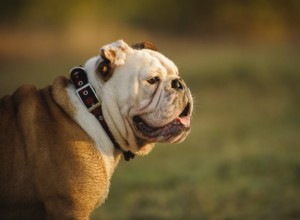
잉글리시 불독으로도 알려진 귀엽고 사랑스러운 불독은 가장 잘 알려진 개 중 하나입니다. 이 품종이 한때 거의 멸종했다고 믿기 어렵습니다. 그러나 이 통통하고 주름진 얼굴의 개 품종은 사람들의 마음을 사로잡았고 미국에서 가장 인기 있는 개 중 하나입니다. 불독을 기르는 데는 상당한 노력과 돈이 필요하지만, 이 개를 가족이라고 부르는 사람들은 평온하고 게으르며 종종 코믹한 동반자에게 보상을 받습니다. 사랑하는 불독을 적절하게 돌보는 데 필요한 역사적 기원, 재미있는 사실 및 정보를 알아보십시오. 역사와 기원 불독 품종의

얼마 전까지만 해도 침술, 지압, 카이로프랙틱, 마사지 요법, 동종 요법이 일부 개 주인과 많은 수의학 시설에서 경멸과 의심으로 여겨졌습니다. 오늘날, 이러한 치유 양식은 전통적인 서양 수의학의 실행 가능한 동반자로 널리 받아들여지고 존경받고 있습니다. 동양과 서양이 협력 관계를 형성함에 따라 더 많은 수의사들이 수의과 대학에서 배운 의료 기술 및 프로토콜과 함께 이를 사용하기 때문에 보완 요법이라고 하는 경우가 많습니다. 지속적인 파행, 소화기 질환, 훈련 및 행동 문제, 기타 여러 불가사의한 송곳니 퍼즐과 같은 전통적인 수의학