랜디 키드, DVM, PhD 작성 서양 의학의 역학 이론은 신체의 관절을 단순히 뼈의 지렛대 작용이 신체의 움직임을 가능하게 하는 해부학적 부위로 간주합니다. 그러나 관절은 해부학적으로, 기계적으로, 기능적으로 이보다 훨씬 더 복잡합니다. 그리고 어떤 관절에 질병이 존재하면 그 결과는 국소적뿐만 아니라 몸 전체를 완전히 무력화시킬 수 있습니다. 관절을 다양한 기능을 가진 신체 기관 시스템의 또 다른 하나로 보면 동물의 전반적인 웰빙의 전체적인 균형에서 관절의 중요성을 더 잘 알 수 있습니다.

우선, 관절은 주변 근육, 인대, 힘줄, 신경, 충격 흡수 장치(척추 디스크 및 관절 반월판), 윤활제를 생성하는 활막의 지지 캐스트 없이는 아무 기능도 할 수 없다는 사실을 인식하는 것이 중요합니다. 또한 관절의 기능적 표면에는 혈액과 림프관이 제대로 공급되지 않고 튼튼한 관절낭을 통해 다른 조직과 분리되어 있습니다. 이러한 순환과 분리의 부족은 관절 내의 모든 질병 과정의 치유를 훨씬 어렵게 만듭니다. 관절의 일부 기능(움직임 생성 레버로서의 기계적 기능에 추가)에 대한 간략한 목록은 다음과 같습니다. • 안정성. 관절을 둘러싸고 있는 관절 안정화 구조에는 인대(뼈에서 뼈로 연결하는 섬유 조직), 힘줄(근육을 뼈에 연결하는 근육의 섬유 끈 모양 확장) 및 근육이 있습니다. 엉덩이를 둘러싸고 있는 적합하고 건강한 근육량을 가진 개는 고관절 이형성증 발병으로부터 가장 잘 보호됩니다. • 고유수용성. 인대, 힘줄, 특히 관절을 둘러싸고 있는 작은 근육에는 고유수용성 신경 말단이 풍부합니다. 신경은 한 번에 모든 신체 부위의 위치를 개의 뇌에 전달합니다. 고유 감각 정보는 특히 개가 움직일 때 신체 균형에 매우 중요합니다. • 윤활. 활막(관절낭의 내부 안감)은 윤활제 역할을 하는 미끄러운 물질을 생성하여 관절 표면이 서로에 대해 자유롭게 움직일 수 있도록 합니다. • 충격 흡수. 뼈 끝은 움직이고 무게를 지탱하는 뼈를 위한 완충기 역할을 하는 부드럽고 연골 조직으로 구성되어 있습니다. 또한 일부 관절에는 추가 패딩이 있습니다. 예로는 척추의 척추뼈와 무릎의 반월상 연골 사이의 디스크가 있습니다(무릎의 반월판은 관절 내에 위치한 초승달 모양의 섬유 연골 패드입니다). 측면 및 회전 운동의 "충격"은 관절의 주변 조직인 근육, 힘줄 및 인대에 의해 억제됩니다. • 스프링 액션. 인대와 힘줄에 가해지는 장력은 고무줄을 조이고 푸는 것과 같은 기계적 방식으로 관절 내에서 스프링과 같은 작용을 만듭니다. 또한, 관절 자체의 해부학적 설정은 스프링과 같을 수 있어 압축 중에는 보호를 제공하고 확장 단계에서는 추가 진동을 제공합니다. (여기에 있는 비절을 생각해 보세요. 개가 뛸 때 압축했다가 튕겨져 나갈 수 있는 놀라운 능력이 있습니다.) • 전신 유연성. 관절은 물론 신체의 유연성을 담당하지만, 이 유연성을 움직임(운동)을 통해 능동적이고 지속적으로 활용하고 균형을 유지하는 것이 중요합니다. • 구조적 재정렬. 관절은 골격의 지지 구조를 구조적으로 재정렬하는 주요 부위입니다. 만성 외상 또는 과도한 구조적 압력(해부학적으로 정렬되지 않은 뼈)은 염증 과정과 새로운 뼈 성장을 시작하여 궁극적으로 압력에 반응하는 관절 측면을 따라 뼈 조직의 양을 증가시킵니다. 이 뼈 성장은 종종 고통스럽고 관절을 사용할 수 없게 만들 정도로 고통스러울 수 있습니다. • 통증. 관절 부위에 위치한 통증이 너무 심해 동물이 움직이기를 꺼려할 수 있으며, 관절이 점점 덜 활동적이 됨에 따라 궁극적으로 해당 부위를 움직일 수 없는 관절로 완전히 융합시키기에 충분한 보상 뼈 조직(외골증)을 생성할 수 있습니다. • 면역 기능. 과학자들은 아직 동물 관절의 면역 기능의 정확한 범위를 결정하지 못했지만 면역 체계가 망가지면 관절이 심각하게 영향을 받을 수 있다는 것을 알고 있습니다. 여기서 류마티스 관절염이 우리의 상징입니다. 관절을 더 자세히 평가하면 건강한 전신 면역 체계를 유지하는 데 관절이 중요한 역할을 한다는 것을 알게 될 것이라고 생각합니다. 관절은 결국 모든 신체 움직임(생명 자체를 결정하는 요소 중 하나)에 관여합니다. 그리고 그들은 신체에서 가장 오래 사는 조직(뼈)과 지속적으로 접촉하고 있습니다. 관절 해부학 간단히 정의하면 관절은 두 개 이상의 뼈가 서로 연결되어 해당 신체 부위의 움직임을 초래하는 지레를 생성하는 부위입니다. 관절이 이 관절을 만드는 방법에는 여러 가지가 있으며 이러한 차이로 인해 관절이 다양하게 분류됩니다. 가장 일반적인 분류는 조인트의 구조적 기하학에 해당합니다. 그 결과 "볼과 소켓" 관절(예:엉덩이) 및 "경첩" 관절(예:사람의 손목과 유사한 개의 손목 관절)과 같은 용어가 생성됩니다. 힌지 조인트는 일반적으로 도어 힌지처럼 한 방향으로만 움직입니다. 진정한 볼 및 소켓 조인트는 전방, 후방, 측면, 내측, 회전 등 모든 방향으로 움직일 수 있습니다. 그러나 일부 볼 및 소켓 조인트는 조인트를 둘러싼 구조로 인해 전체 움직임이 다소 제한됩니다. 주변 구조물 많은(전부는 아니지만) 관절은 관절의 내부 작용을 효과적으로 보호하고 부상과 감염에 대한 장벽을 만드는 두꺼운 섬유질 캡슐로 둘러싸여 있습니다. 관절낭의 내벽인 활막은 관절의 윤활제 역할을 하는 두꺼운 젤라틴성 액체를 생성합니다. 관절과 뼈 끝의 연골 조직에 필요한 대부분의 영양소는 활액에서 나옵니다. 연골과 관절 표면은 혈관에서 제대로 공급되지 않기 때문에 중요한 고려 사항입니다. 활막 조직은 외상과 감염에 반응합니다. 그 결과 염증 성분의 생산 증가와 함께 조직 두께가 증가합니다. 이 염증성 활액 반응은 바늘 생검을 통해 평가할 수 있습니다. 힘줄은 근육을 뼈에 부착시키는 근육의 섬유성 확장입니다. 근육이 수축할 때 관절의 "지레"를 가로질러 부착된 뼈를 움직입니다. 인대는 관절을 가로질러 뼈에서 뼈로 연결됩니다. 그들의 주요 기능은 관절을 안정화시키는 것입니다. 인대와 힘줄은 모두 튼튼한 구조로, 건강할 때 관절이 정상적으로 노출될 수 있는 몇 배의 힘을 견딜 수 있을 만큼 충분한 강도(거의 뼈 자체만큼)를 갖습니다. 인대와 힘줄은 주로 섬유질 조직과 연골 조직으로 구성되어 있지만 둘 다 약간의 신축성 조직을 포함하여 일정량의 스트레칭을 허용합니다. 근막은 힘줄과 인대를 둘러싸고 있는 섬유질 결합 조직으로, 큰 근육 덩어리를 작은 근육 "배"로 나눕니다. 근막은 또한 근육을 통과하는 혈관을 보호합니다. 사실, 혈관을 둘러싸고 있는 근막 담요의 수축과 이완은 개가 활동적일 때마다 힘줄을 통해 관절로 가는 혈류를 향상시키는 2차 혈액 펌프 역할을 합니다. 강아지가 충분한 운동을 하도록 하는 또 다른 이유입니다. 관절을 둘러싸고 있는 근육은 또한 안정성과 고유수용감각의 중요한 구성요소입니다. 근육이 잘 맞을수록 관절이 더 안정적입니다. 운동이 관절 주변의 작은 근육에 고유수용성 신경 중추를 발달시키는 데 도움이 되므로 건강한 사람에게 더 나은 균형 감각을 제공한다는 증거가 있습니다. 관절 기능 관절의 적절한 기능은 다음과 같은 여러 요인에 따라 달라집니다. • 관절 표면의 적절한 정렬을 생성하는 해부학 – 관절이 원래 이동하려는 방향으로 움직일 수 있도록 하는 골격 정렬 • 다음과 같이 유연성을 제공하는 주변 조직 안정성 • 비교적 매끄럽고 쉽게 움직일 수 있도록 윤활 처리된 관절 표면 • 관절을 뇌에 연결하고 관련된 신체 부위의 정확한 위치를 전달하는 기능하는 고유수용 신경계 • 균형 잡힌 전신 면역 경미한 감염이나 부상을 막기 위해 면역 반응을 유지할 수 있고 과민성 면역 반응을 일으키지 않고 그렇게 할 수 있는 시스템입니다(예:류마티스 관절염에서 볼 수 있음). 조인트 및 조인트 표면 관절의 생리학에 대한 최근 연구들이 많이 있었습니다. 아마도 우리가 이제 관절이 개와 인간 모두에서 질병의 주요 부위임을 인식하기 때문일 것입니다. 거의 7천만 명에 가까운 사람들이 관절염이나 어떤 형태의 만성 관절 통증으로 고통받고 있으며 관절 질환, 특히 만성 질환은 전체론적 수의사들이 보는 가장 큰 질환일 수 있습니다. 관절 생리가 양호하면 뼈가 서로 접촉하는 매끄러운 표면이 쉽게 관절을 이루고 뼈의 무게 지지력이 충격에 의해 완충됩니다. 관절의 기능적 건강은 윤활 물질의 적절한 생산과 결합된 연골 재생 및 퇴행의 균형에 의해 달성됩니다. 뼈의 연골 말단은 대부분이 물(전체 기질의 약 65~80%)이며 기질의 대부분은 콜라겐과 프로테오글리칸의 혼합물입니다. 혼합물에는 연골 조직의 복구 및 조절을 담당하는 세포인 소수의 연골 세포가 있습니다. 콜라겐은 다양한 형태로 존재하고 다양한 기능을 수행하는 1차 결합 조직입니다. 그것은 몸 전체에 걸쳐 접착제 또는 아교와 같은 물질처럼 작용하고 구조를 유지하는 데 도움이 되며 연골에서 프로테오글리칸을 제자리에 고정하는 프레임워크를 제공할 뿐만 아니라 탄력과 충격 흡수를 제공합니다. 프로테오글리칸은 당과 단백질로 구성된 복잡한 분자입니다. 그들은 콜라겐 섬유와 상호 연결되어 연골을 탄력있게 만들어 신축성이 있고 제자리로 돌아갈 수 있습니다. 프로테오글리칸은 또한 스펀지처럼 작용하여 물을 가두어 관절의 지속적인 움직임에 필요한 유연성을 연골에 제공합니다. 프로테오글리칸은 코어 단백질에 연결되어 관절 치유에 사용되는 중요한 구성요소인 글리코사미노글리칸(GAG)을 형성합니다. 마모의 형태로 또는 감염 유도 및/또는 면역 매개 퇴행 과정의 결과로 과도한 요구가 있으면 뼈의 연골이 침식될 수 있습니다. 다시 한 번, 우리는 관절이 과도하게 마모되고 찢어지는 가장 일반적인 이유를 만들었습니다. 종의 부자연스러운 골격 구조를 가진 개는 관절이 비정상적으로 마모되고 찢어지기 쉽습니다. 과도한 관절 침식은 염증 반응을 촉진하여 활막이 두꺼워지고 백혈구 및 기타 염증 산물이 방출됩니다. 연골의 미란이 진행되면서 관절 표면이 거칠어지고 결국 동물은 통증을 느끼기 시작할 수 있습니다. 추가 침식은 연골의 보호층을 제거하여 뼈가 뼈에 문지르게 할 수 있습니다. 신체의 치유 메커니즘은 뼈에 대한 뼈를 과도한 구조적 스트레스로 해석하고, 신체는 스트레스에 대응하기 위해 더 많은 뼈를 생성함으로써 반응합니다. 이 "새로운" 뼈는 종종 관절을 둘러싸고 있는 뼈 조직의 무질서한 덩어리(외골)로 형성되며, 이러한 뼈 증식은 부드러운 조직이 뼈에 문지름에 따라 더 많은 통증(및 더 많은 염증)을 유발합니다. 그 과정을 멈출 수 있는 것이 없으면 진행되고 만성화되며 점차 악화됩니다. 손상된 관절 조직의 복구는 유발 원인이 제거되거나 제거된 후에 시작할 수 있습니다. 그런 다음 뼈 끝에 위치한 소수의 연골 세포가 새로운 연골 조직을 (천천히) 추가하기 시작할 수 있습니다. 새로운 연골 조직 합성의 핵심 구성 요소는 글루코사민이 기본 빌딩 블록인 글리코사미노글리칸(GAG)의 생성입니다. 힘줄 및 인대 힘줄은 근육을 뼈에 연결하는 끈 모양의 근육 확장입니다. 이 부착물을 통해 힘줄이 인접한 뼈를 움직입니다. 이 지렛대 작용은 힘줄이 미끄러지고 미끄러지는 능력에 달려 있습니다. 이는 힘줄을 둘러싸고 있는 미끄럽게 표면이 된 외피에 의해 가능해진 능력입니다. 힘줄 외피는 새로운 조직 성장과 조직 분해의 지속적으로 균형 잡힌 과정에 의해 기능을 유지하며, 새로운 조직은 항상 미끄러워야 하는 힘줄의 요구에 맞춰 정렬됩니다. 여기 문지름이 있습니다. 힘줄이 심하게 손상된 경우 신체는 흉터를 만들어 이를 복구하려고 시도하며 흉터의 유일한 목적은 손상된 눈물 끝을 다시 결합하는 것입니다. 찢어진 힘줄이 치유됨에 따라 흉터는 힘줄의 정상적으로 미끄러운 기능을 되돌리지 않고 끝이 융합될 가능성이 높습니다. 정상적인 힘줄 기능을 유지하는 데 도움이 되는 여러 가지 수술 방법이 있지만 주의할 점은 수술 시도는 즉시(몇 시간 이내에) 이루어져야 하며 외과의는 관절 수리에 능숙해야 한다는 것입니다. 초정밀 관절 운동의 필요성은 일반적으로 사람(특히 사람의 손가락)에서만큼 개에서 그리 크지 않으며 적절한 기능은 일반적으로 "정상적인" 수술 절차 후에 유지될 수 있습니다. 힘줄 수리에는 약 6주가 소요됩니다. 처음 3주 동안 힘줄의 반대쪽 끝은 움직이지 않도록 해야 합니다. 다음 3주 동안 힘줄은 미끄러짐을 제공하기 위해 힘줄 외피를 추가하는 것을 포함하는 리모델링을 허용하기 위해 약간의 움직임(덜 격렬한 외부 부목을 통해)이 필요합니다. 완전한 치유에는 1년 이상이 걸릴 수 있으며 완전히 치유된 힘줄은 원래의 90% 정도만 강해질 것입니다.

인대 치유는 예측할 수 없습니다. 일부는 저절로 치유됩니다. others are almost impossible to repair, even with the best of surgical techniques. For these problem ligaments, techniques have been developed to use transplanted fibrous tissues to repair the damage. Another potential problem arises when bone fragments or particles of cartilage flake off from areas of joint inflammation, and they become abrasive “floaters” that add to a joint’s pain and inflammatory process. Usually, the only cure for these floaters is to have them surgically removed. Whenever you suspect severe trauma to a joint – for example, when your dog experiences a sudden onset of limping, exhibits obvious pain when moving, or refuses to walk, climb stairs, jump up on or down from furniture, or rise from a sleeping position – see a vet as soon as possible, and consider getting a second opinion from a board certified veterinary surgeon. Diseases of joints and surrounding tissues • Osteochondrosis is a disturbance in the formation of normal cartilaginous tissue; it’s considered a congenital or inherited disease. Immature articular cartilage separates from the underlying bone and floats free in the joint cavity, causing pain, inflammation, and eventually excess bone growth within and around the joint. The disease may affect the shoulder, elbow, stifle, or hock joints. Osteochondrosis typically develops in large breed dogs, and the lesions occur during the maximal growth phase of the skeleton – when the dog is four to eight months of age. Limiting the growth phase of large breed dogs may help prevent the disease. Floating bits of cartilage (also called “joint mice”) need to be removed surgically, and joint fluid modifiers such as glucosamine may also help with repair and prevent further damage to the articular surfaces. Acupuncture may also speed healing. Prognosis for recovery is excellent for shoulders, good for the stifle, and fair for the elbow and tarsal (hock) joints. • Elbow dysplasia is a generalized term that describes several entities, all of which result in abnormal elbow joint function (some of which seem to be genetic). Elbow problems typically develop in young, large, rapidly growing dogs – affected animals demonstrate abnormal bone growth, joint stresses, or cartilage development. The joint is painful, causing the dog to limp, and radiographs are often needed to confirm the various conditions. Treatment is the same as for osteochondrosis, discussed above. • Hip dysplasia may be the biggest joint problem to confront dog owners and breeders in this country. This multifactorial disease affects a high number of dogs, and we don’t yet have a good handle on what causes it, nor what is an effective cure. Further, there is a definite genetic component to hip dysplasia, but the genetics aren’t clear-cut. Some dogs that aren’t supposed to be affected (according to statistical probability) are, and visa versa. While hip dysplasia is far too vast a topic to cover in this article, the following observations may be helpful. Excessive growth, exercise, nutrition, and hereditary factors all affect the occurrence of the disease. Dogs who grow too rapidly for the amount of muscle mass surrounding their hip joints are more prone to joint laxity, which in turn allows for excess movement within the joint. As the joint becomes increasingly unstable, its excessive movements lead to inflammation and ultimately degenerative joint changes – fibrosis, bony growths around the joint, flattening of the femoral head and the acetabulum (the socket joint of the hip that the “ball” of the femoral head fits into), and possibly subluxation or luxation of the femoral head. The changes of hip dysplasia tend to be progressive; however, dogs do not always have symptoms that correspond to the severity of the changes evident on radiographs. Some dogs are practically immobile with only slight changes; others might have severe changes but appear symptom-free. Dogs with symptoms have varying degrees of lameness that tend to get worse with exercise. The first sign of hip dysplasia is often a dog’s reluctance to climb stairs or difficultly when getting up or lying down. Some dogs with hip dysplasia may exhibit a rabbit-like, hopping gait when running. Radiographs help determine the amount of physical damage to the joint, but don’t always correlate with the dog’s symptoms. Prevention efforts are primarily aimed toward identifying potentially affected individuals (with screening X-rays and palpation) and removing them from the breeding pool. There is new interest in the potential of gene mapping. Each of the methods has its own strengths and shortcomings. Look for a board certified veterinary radiologist to do any final evaluations. Conventional treatments include pain relief and various forms of surgery. I’ve found alternative medicines to be especially helpful for hip dysplasia; I use a combination of acupuncture and chiropractic along with nutritional and nutriceutical remedies and possibly herbal support. • Septic or infectious arthritis can be from penetrating trauma (including surgery) or from a spread of infection from other parts of the body. Pain and swelling are common symptoms of infected joints. A needle biopsy of the joint may reveal increased numbers of white blood cells and possibly the instigating microorganism; X-rays may indicate inflammatory response and/or bony changes. Due to the lack of an abundant blood supply to the area, joint infections often require high doses of the specific antibiotic indicated for the microorganism involved. • Immune-mediated arthritis is the consequence of secondary deposits of immune complexes within joints, usually affecting all the joints of the body. These immune complexes cause an inflammatory response that may erode the joint surface (rheumatoid arthritis) or be nonerosive as with systemic lupus erythematosus (SLE). Joints swell and are painful, the dog becomes lame, and he may run a fever and refuse to move or eat. Treatments are aimed at eliminating the pain and rebalancing the immune system. The immune-mediated diseases are another area where I’ve had especially good results using alternative medicines. Most of the alternative approaches (especially acupuncture, homeopathy, and herbal remedies) can help balance the immune system while the remedies are being directed toward specific areas of disease. • Neoplastic arthritis is rare – fortunately, because it tends to be an aggressive neoplasia; the usual recommendation is to amputate the limb. • Trauma to joints is not uncommon. It may present itself in several ways:Joint fractures may occur in any joint, but they are most common in immature animals. They typically occur within the joint, at the growth plate (physis) of the bone, which is its weakest point. The goal for treating a joint fracture is to bring the fractured ends back together and to hold them there until healing takes place. Ligament tears and ruptures are also common. Pain and swelling are evident, and the intensity of the symptoms depends on the extent of the tear. The anterior/cranial and posterior/caudal cruciate ligaments help stabilize the knee joint by crossing from lateral to medial (thus the term “cruciate”) and spanning across the joint from the femur to the tibia. Rupture of the anterior cruciate ligament is a fairly common occurrence. It is caused by excessive trauma, weakened ligaments from immune-mediated causes, and/or poor conformation (straight-legged dogs). Diagnosis of a cruciate ligament is made through palpation (to discern the amount of abnormal movement of the knee) and x-rays. Some dogs recover without any treatment; others respond to weight reduction, immobilization of the joint, acupuncture, and physical therapy; still others will require surgical repair. Surgery typically grafts a tendon from another part of the dog’s body to act as a replacement for the one torn. • Traumatic dislocation of the hip, or hip luxation, is often the result of being hit by a car. It results in lameness, pain when the leg is manipulated, and a shortened affected limb. Treatment either involves nonsurgical manipulation of the limb to reposition it, followed by the use of slings to maintain it in the joint, or surgical stabilization using pins or sutures. Treatment overview Treating any ongoing arthritic problem, no matter its cause, involves the following:• Halting the initial reason that caused the inflammatory process • Repairing the damage already present • Returning the joint (as much as possible) to normal function; without function, the joint will ultimately fuse • Enhancing the healing process. Repair of the damaged surface of a joint depends on several factors, including:• Removal of the inciting cause. Examples include removing exostoses and joint floaters, eliminating infection, or rebalancing the immune system in the case of immune-mediated arthritis. • Return to a more normal joint movement (if possible). • Pain control. Returning to normal joint movement usually requires some easing of existing pain, either via acupuncture, chiropractic adjustments, or herbal (or other) remedies. And, a joint will remain normal only if its functional way of moving has returned to near-normal. • Provide the necessary basics for healing, using nutrients and nutriceuticals Natural medicines for joints I consider chiropractic and acupuncture as the one-two punch for any problem involving the musculoskeletal system; both are especially effective for treating joint conditions. Chiropractic is used in an attempt to return the joint to its normal function – a necessary prerequisite for any long-term healing of the joints. Acupuncture is a proven therapy for alleviating pain and enhancing the immune system. Plus, it may offer a necessary “energetic” to enhance healing. Homeopathic remedies, especially when used in the classical way, may be curative for joint diseases, but I generally consider them to be more helpful as an acute therapy for pain. Remedies that I have found to be effective for my patients include:Arnica, used early-on for acute pain; Rhus tox for the “rusty gate” syndrome – the limp that gets better with use; Byronia for the limp that gets worse with movement; Hypericum for pain; and Ruta for deep pain. Massage and physical therapy can be vital therapies to help in the healing process. These methods help eliminate pain, and physical therapy, in particular, helps return the joint’s normal function by helping it move through its normal range. There are dozens of herbs that can be helpful for treating joint diseases, and they can be especially helpful for providing nontoxic pain relief and for balancing the immune system. In addition, there are some herbs that have been used to treat specific joint problems. Check with an herbalist who has some experience using herbs to treat animals for the appropriate herbs and their dosages. [Editor’s note:Also see Dr. Kidd’s book, Dr. Kidd’s Guide to Herbal Dog Care.] Nutrients and nutriceuticals. There has been much recent buzz about using nutrients and nutriceuticals for joint healing, and the interest has been for good reason – many, if not most, animals with joint conditions respond to varying degrees after a month or so of treatment. There are several of the glycosaminoglycans (GAGs) and proteoglycans that have been used. The most popular are glucosamine (the basic building block for cartilaginous tissue) and chondroitin sulfate, which prevents other body enzymes from degrading the building blocks of joint cartilage. Methylsulphonylmethane (MSM) is another substance that is often added to “joint-repair” mixtures. It supplies needed sulphur molecules and seems to provide additional pain relief. There are a variety of these products available commercially, and even some of today’s commercial dog foods contain them (although probably not in amounts that could possibly be therapeutic). My own experience would indicate that it will take your dog at least 30 days to respond to any of the nutriceuticals mentioned. Further, one product does not fit all; in my experience, some dogs respond to one product and not others, and visa versa. If one product doesn’t seem to be working after a few months trial, first try increasing the dosage for a month or so. If that doesn’t work, try another product. In every case I can think of, we have eventually come up with a product that seems to be helpful. Recent evidence indicates that the Omega-3 fats are beneficial for helping with the joint healing process. Manganese is needed for healthy cartilage formation and it is used in several enzymatic processes in the body. Supplemental vitamin C (especially in the form of sodium ascorbate) is also beneficial for tissue healing. For dosages and method of application of the nutriceuticals and nutrients, check with your holistic veterinarian. Also With This Article “Joint Supplements for Dogs” “Identifying Arthritis in Dogs” -Dr. Randy Kidd earned his DVM degree from Ohio State University and his PhD in Pathology/Clinical Pathology from Kansas State University. A past president of the American Holistic Veterinary Medical Association, he’s author of Dr. Kidd’s Guide to Herbal Dog Care and Dr. Kidd’s Guide to Herbal Cat Care.
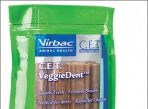
호주에서 리콜된 VeggieDent Chews 문제를 일으키는 것으로 의심되는 조사 6월에 Virbac은 수의학자들이 강아지의 신장 질환 발병과 이러한 씹는 음식 사이의 연관성을 발견한 후 예방 조치로 호주의 VeggieDent Chews for Dogs를 회수했습니다. 이유는 발견되지 않았으며 씹는 것이 판코니 유사 증후군을 유발한다는 증거도 아직 없습니다. 츄어는 올해 3월 호주에 도입됐다. 베트남에서 제조되며 옥수수, 전분, 글리세린, 콩, 쌀, 효모, 소르비톨, 옥수수 유도체 및 물로 만들어집니다. 288 Veggi
Iverhart Plus 리콜 무게가 75파운드 이상인 개는 완전히 보호되지 않을 수 있습니다. 8월 13일 Virbac Animal Health, Inc.는 심장사상충 예방제인 Iverhart Plus® 맛 츄어블 정제의 일부를 리콜한다고 발표했습니다. 일상적인 안정성 테스트 결과 이 로트에는 체중이 75파운드 이상인 개에게 효과적인 이버멕틴이 더 이상 함유되지 않은 것으로 나타났습니다. Iverhart Plus Large(51-100파운드)의 로트 번호 090093 및 090095가 리콜되었습니다. 소형견을 위한 세 번째